Welcome to the Amira-Avizo Software Use Case Gallery
Below you will find a collection of use cases of our 3D data visualization and analysis software. These use cases include scientific publications, articles, papers, posters, presentations or even videos that show how Amira-Avizo Software is used to address various scientific and industrial research topics.
Use the Domain selector to filter by main application area, and use the Search box to enter keywords related to specific topics you are interested in.

Protocols for Generating Surfaces and Measuring 3D Organelle Morphology Using Amira
High-resolution 3D images of organelles are of paramount importance in cellular biology. Although light microscopy and transmission electron microscopy (TEM) have provided the standard for imaging cellular structures, they cannot provide 3D images.
However, recent technological advances such as serial block-face scanning electron microscopy (SBF-SEM) and focused ion beam scanning electron microscopy (FIB-SEM) provide the tools to create 3D images for the ultrastructural analysis of org... Read more
Edgar Garza-Lopez, Zer Vue, Prasanna Katti, Kit Neikirk, Michelle Biete, Jacob Lam, Heather K. Beasley, Andrea G. Marshall, Taylor A. Rodman, Trace A. Christensen, Jeffrey L. Salisbury, Larry Vang, Margaret Mungai, Salma Ash Shareef, Sandra A. Murray, Jianqiang Shao, Jennifer Streeter, Brian Glancy, Renata O. Pereira1, E. Dale Abel, and Antentor Hinton, Jr.
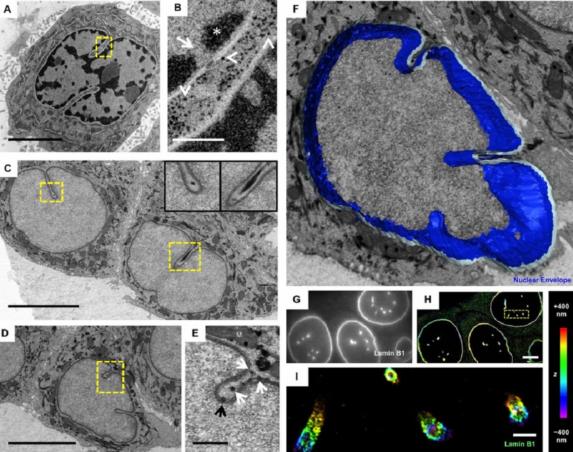
The importance of context in regulation of gene expression is now an accepted principle; yet the mechanism by which the microenvironment communicates with the nucleus and chromatin in healthy tissues is poorly understood. A functional role for nuclear and cytoskeletal architecture is suggested by the phenotypic differences observed between epithelial and mesenchymal cells…
Read more
Danielle M. Jorgens, Jamie L. Inman, Michal Wojcik, Claire Robertson, Hildur Palsdottir, Wen-Ting Tsai, Haina Huang, Alexandre Bruni-Cardoso, Claudia S. López, Mina J. Bissell, Ke Xu, Manfred Auer
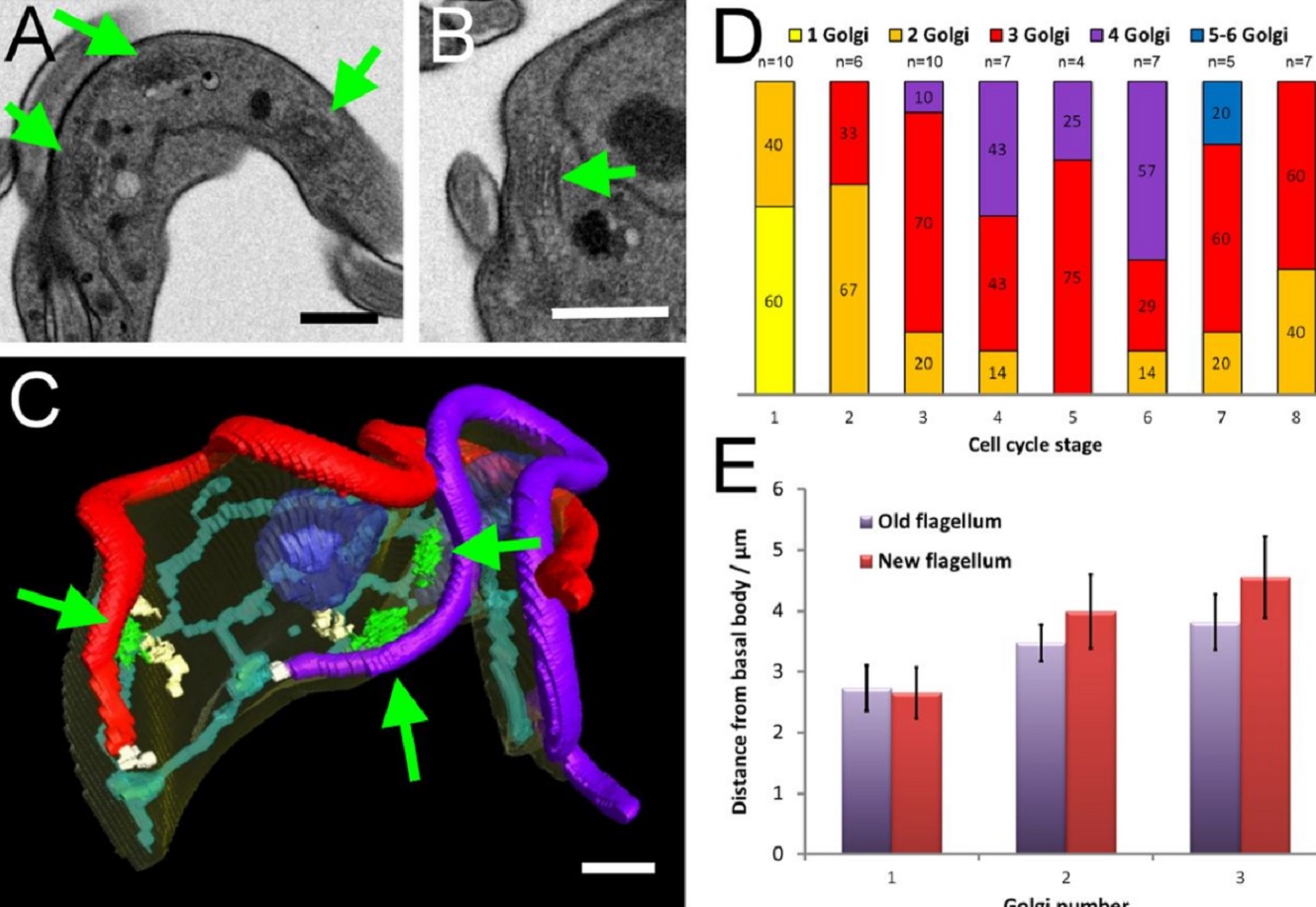
The major mammalian bloodstream form of the African sleeping sickness parasite Trypanosoma bruceimultiplies rapidly, and it is important to understand how these cells divide. Organelle inheritance involves complex spatiotemporal re-arrangements to ensure correct distribution to daughter cells…
Read more
Louise Hughes, Samantha Borrett, Katie Towers, Tobias Starborg, Sue Vaughan
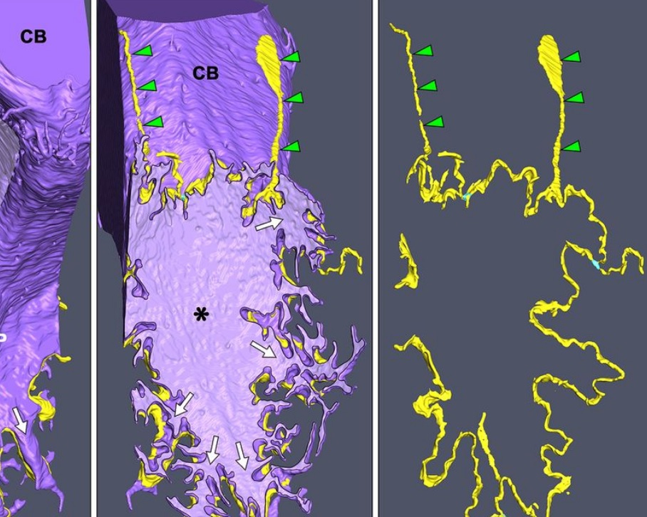
Morphological process of podocyte development revealed by block-face scanning electron microscopy
Podocytes present a unique 3D architecture specialized for glomerular filtration. However, several 3D morphological aspects on podocyte development remain partially understood because they are difficult to reveal using conventional scanning electron microscopy (SEM). Here, we adopted serial block-face SEM imaging…
Read more
Koichiro Ichimura, Soichiro Kakuta, Yuto Kawasaki, Takayuki Miyaki, Takahiro Nonami, Naoyuki Miyazaki, Tomoyo Nakao, Sakiko Enomoto, Shigeo Arai, Masato Koike, Kazuyoshi Murata, Tatsuo Sakai